Young Fabry Disease Patients Show Brain Lesions in the Absence of Clinical History of Stroke

Children and adolescents with Fabry disease who have no clinical history of stroke, show asymptomatic brain lesions, according to a follow-up neuroimaging study. Early detection of these lesions could make it easier for clinicians to begin treatment.
The study, “Brain MRI findings in children and adolescents with Fabry disease” was published in the Journal of Neurological Sciences.
In Fabry disease, accumulation of a type of fat called globotriaosylceramide (Gb3) — caused by a deficiency in the alpha-galactosidase A, an enzyme encoded by the GLA gene — and other fat molecules, leads to the development of cerebrovascular disorders (stroke and others), as well as cardiac and renal complications.
Both ischemic stroke (when an artery to the brain is blocked, stopping delivery of oxygen and nutrients) or hemorrhagic stroke (abnormal accumulation of blood that compresses the surrounding brain tissue) can occur at an early age in Fabry patients.
It is estimated that 1% to 2% of all strokes in the general population aging from 18 to 55 may be due to Fabry disease.
The study authors previously reported the presence of asymptomatic ischemic (44.4%) and microbleeds (11%) brain lesions in a group of Fabry adult patients without a history of stroke.
In this follow-up study, researchers evaluated the presence of white matter lesions (WML) and hemorrhagic lesions in young Fabry patients, as the occurrence of such lesions in children and adolescents with Fabry disease, without a history of stroke, has not been commonly reported.
WML is a clinical condition commonly seen in elderly people that results from loss of myelin, a fatty material that protects nerve fibers and allows communication between nerve cells.
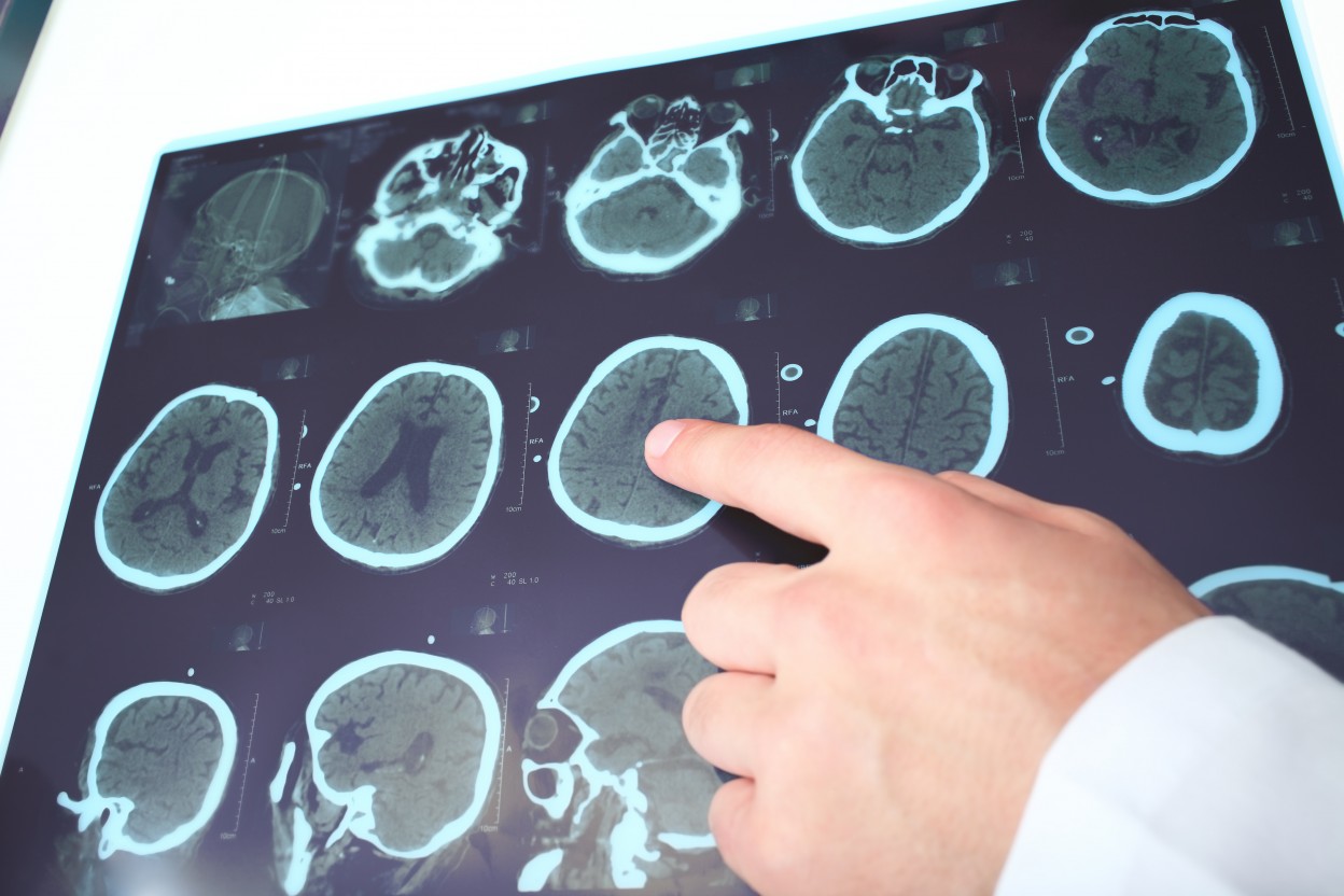
The study involved the review of brain magnetic resonance imaging (MRI) — a technique that allows detailed pictures of the brain — of 44 children and teenagers, 20 of whom were boys, aging from 7-21 years (mean age of 14.6), from 13 different families with Fabry disease and no prior history of stroke.
Forty-six healthy children and adolescents were used as controls (36 females, mean age 14.1 years).
Most (90.9%) patients presented signs or symptoms of Fabry disease, with chronic pain (neuropathic pain) being the most frequent.
Of the 44 Fabry patients, seven children (15.9%) exhibited brain MRI evidence of asymptomatic WML (of which five were girls). These numbers were significantly higher when compared to the control group, where only three children (6.5%) showed evidence of WML.
Brain abnormalities were more frequent in heterozygous Fabry females (often regarded as being asymptomatic or having a less severe form of the disease), which according to the authors indicates that “in this disorder both females and males could be affected similarly.” Heterozygotic means that an individual has two different copies of a particular gene.
Contrary to what happens with adult Fabry patients, none of the young patients participating in this study presented signs of brain hemorrhages.
“Fabry disease (…) should routinely be included in the differential diagnosis of both male and female patients with symptomatic and asymptomatic brain lesions in children and adolescents,” the researchers wrote, adding that “the early identification of WML may foster the decision to initiate treatment.”


