
Absence of Lesions in Two Specific Brain Regions May Help Distinguish Fabry Disease from MS, Study Shows
Written by |


The absence of white matter lesions (WMLs) in two specific regions of the brain may help physicians distinguish Fabry disease from multiple sclerosis and make an accurate diagnosis, a study has found.
The study, “Absence of infratentorial lesions in Fabry disease contributes to differential diagnosis with multiple sclerosis,” was published in the journal Brain and Behaviour.
White matter forms the largest and deepest part of the brain and the superficial part of the spinal cord. It is made up of nerve cell axons (fibers), which are responsible for the transmission of nerve signals between the different areas of the brain and spinal cord.
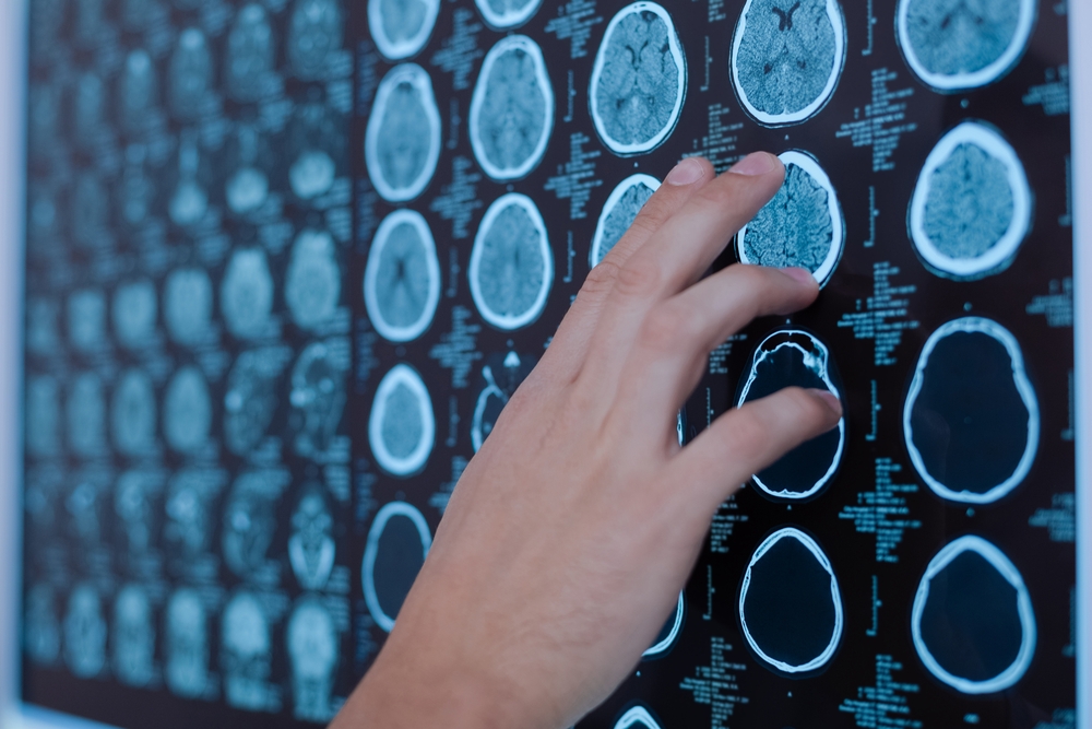
WMLs, commonly detected on brain magnetic resonance imaging (MRI) scans, can disrupt nerve cell communication and cause neurological problems.
Fabry disease often causes damage to the central nervous system (brain and spinal cord), including WMLs and strokes. The presence of WMLs and the wide range of symptoms in these patients can lead to a misdiagnosis of multiple sclerosis (MS), a neurodegenerative disease characterized by WMLs.
That is why identifying diagnostic measures — including those detected in MRI scans — that could help distinguish these two conditions is key for early and correct diagnosis, and implementation of appropriate treatment.
While multiple sclerosis is known to affect mainly specific areas of the brain (including the corpus callosum, temporal lobes, brainstem, and cerebellum), which brain areas are mostly affected in Fabry disease remains unexplored.
Recent evidence has shown that WMLs in the corpus callosum — the largest white matter structure in the brain, which connects the two cerebral hemispheres — are much less frequent in Fabry disease patients than in multiple sclerosis patients, suggesting this could be used as a diagnostic measure to distinguish the two conditions.
Also, multiple sclerosis patients often have lesions in the infratentorial region, comprising the cerebellum (controls movement coordination) and the brainstem (responsible for respiratory, heart, motor and sensory functions), but whether this region is frequently affected in Fabry disease remains unclear.
Researchers at the University of Naples Federico II evaluated the frequency of infratentorial lesions in Fabry disease patients and whether this measure (alone or in combination with corpus callosum lesions) could be used as a diagnostic tool to differentiate Fabry disease from MS.
The team retrospectively analyzed MRI scans of 144 Fabry disease patients (mean age of 42 years) and 136 patients with MS (mean age of 38 years).
While all multiple sclerosis patients showed WMLs, only 71 (49.3%) patients with Fabry disease had the presence of WMLs.
The results also confirmed that WMLS in the corpus callosum were substantially more frequent in patients with MS (89.0%) than in Fabry patients (5.6%). The same was found to be true for infratentorial lesions, with 119 (87.5%) MS patients versus only 17 (11.8%) patients with Fabry disease showing WMLs in this region.
When researchers considered the simultaneous presence of WMLs in both areas they found that only four (2.8%) patients with Fabry disease had lesions on both brain regions, compared to 112 (82.4%) patients with MS.
Additional analysis showed that the best overall diagnostic performance was achieved when combining the presence of lesions in both the corpus callosum and the infratentorial region. This was significantly superior to the diagnostic performance obtained using the presence of lesions in only one of the regions, as well as the presence of lesions in at least one of the two regions.
While these results suggest that infratentorial lesions are much less frequent in Fabry disease than in MS, their role as a diagnostic tool to differentiate the two conditions is “less significant compared to the evaluation of CC [corpus callosum], which apparently remains the best indicator for differential diagnosis between FD [Fabry disease] and MS,” researchers wrote.
They also hypothesized that the development of lesion probability maps specific for each condition could have an important diagnostic value.
The team also noted that the appearance of these lesions was different between Fabry and MS patients, suggesting that not only the location, but also the morphology, of WMLs could help physicians “avoid misdiagnosis, providing subsequent correct and prompt treatment options.”


Leave a comment
Fill in the required fields to post. Your email address will not be published.